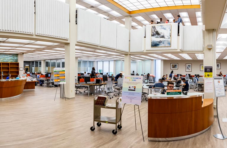
OVERVIEW: EVALUATION

FREQUENCY OF EVALUATIONS

  1. Initial - Evaluation is the first and most important step and should be done as early as possible following injury to provide a baseline against which future changes can be measured. If a good evaluation is not performed soon after the injury, commonly occurring improvements or deteriorations in a patient's neurological status might not be noted. During the initial evaluation, the therapist should, at all times:
    • Involve the patient as much as possible in the evaluation. This gives the patient an active role in the therapeutic program at the outset
    • Relate to the patient as an equal, and not someone who is helpless
    • Provide the patient with complete and accurate information, so the patient can ask questions and learn as much as possible about his/her injury

  2. Periodic - Following the first comprehensive evaluation, therapists should continue to routinely observe changes in the patient's status. More formal and complete evaluations should be performed periodically.

  3. Discharge - A comprehensive examination should be performed upon discharge, for future use by health care providers, to give the patient a full description of his/her status and identify areas for future improvement, and to evaluate the efficacy of the physical therapy program.

GENERAL COMPONENTS

  1. Patient Statements - Record patient statements that reflect his/her emotional status, discomforts, goals, and awareness of the injury.

  2. Patient History - From the medical record and the patient interview, include the following in the patient history:
    • Date, level, extent, and cause of the injury
    • Complications or concurrent injuries
    • Changes in neurological status since the injury
    • Medical conditions that exist
    • Medical and surgical treatment since the injury, with an emphasis on techniques of fracture management
    • Lifestyle before and following the injury, with an emphasis on previous employment and living accommodations, and on existing limitations, such as dependence on a respirator
    • Previous hospital stays
    • Rehabilitative therapy and functioning since the injury

  3. Equipment Check - The type, condition, and suitability of equipment owned or rented by the patient, as well as equipment needed by the patient, should be recorded. Arrangements should be made for equipment procurement or repair. Potential obstacles to therapy, such as respirators and skin problems, should be noted.

  4. Home Environment - Following an initial assessment of the environment to which the patient will return upon discharge, a detailed assessment by a health professional should be performed and modifications made prior to discharge.

MOTOR AND SENSORY EVALUATION

  1. Voluntary Motor Function - An accurate and comprehensive baseline muscle test is needed to predict the extent of function that can be recovered with therapy, identify weak areas that require strengthening, define the neurological status of the patient, and permit progress to be documented. The following guidelines for muscle testing should be observed:
    • A complete muscle test should be performed in extremities that have any voluntary motor function. A gross test should be performed in extremities that are innervated above the level of the injury, followed by a complete test where there are weak areas. A representative test of a muscle from each myotome should be performed in an extremity where there is no apparent motor function.
    • To prevent substitution, immobilize other joints and palpate the muscle being tested to be sure that it is the muscle that is contracting (and not another muscle that the patient has learned to use in the place of a muscle that is weak or nonfunctional).
    • Provide the needed stabilization whenever the muscles that normally provide the needed stabilization are weak or nonfunctional (to prevent muscles from testing weaker than they are due to impaired stability).
    • Request that the patient contract and relax each muscle being tested upon command (to differentiate between voluntary muscle function and reflexive contraction or spasticity).
    • Address complications, such as:
      • Orthoses and medical problems, by performing the muscle test in a different position and documenting the change in position
      • Acute injuries in which the spine is not yet stable, by avoiding the test if the contracted muscle produces exertion on the unstable spine
      • Abdominal and back extensor muscles which may not function along their entire length following SCI, by palpating and noting the motion of the umbilicus and then recording the approximate myotome levels of function (rather than a grade for the muscle)

  2. Sensation - Accurate sensory tests are needed to further define the patient's neurological status, further predict the patient's ability to recover function, and identify areas without sensation that are more susceptible to trauma and skin problems. The following guidelines for sensory tests should be observed:
    • Test proprioception in all joints of extremities that are affected by the injury
    • Test pain and light touch in all key sensory areas of the skin identified by the American Spinal Injury Association (ASIA) and designated with a (.) in the following chart. Since the same spinal cord tract carries both pain and temperature, testing for only one is necessary.

  3. Muscle Tone - Muscle tone tests, needed to further define physical status, involve testing deep tendon reflexes and resistance to passive stretch. Spasticity, if present, should be tested, by the clinician moving the patient's extremities through the available range of motion, and graded according to the following "Modified Ashworth Scale for Grading Spasticity":

  4. Range of Motion - ROM tests, needed to further predict the extent of functional recovery that can be expected with therapy, should include a gross test of all joints, tests of specific joints where limitations are present, and tests of the flexibility of the following muscles which impact function: biceps brachii, pectorialis major, long finger flexors and extensors, rectus femoris, hamstrings, and gastrocnemius. The hamstrings should be tested with the patient is a supine position. Stress to all areas of vertebral instability should be avoided.

  5. Functional Capabilities - Perhaps the most important component of the physical therapy evaluation, the functional evaluation should be highly specific and accurate. Therefore, the therapist should personally observe the patient performing activities and should describe the patient's performance in terms of specific distances, terrains, extent of assistance needed if any, time to accomplish, specific equipment needed if any, etc.
    • The following activities should be observed in a functional evaluation:
      • Rolling, coming to sit, and gross mobility on an exercise mat and also on a bed
      • Even (level), uneven (unlevel), and independent transfers, including assistance required and safety of transfer
      • Wheelchair propulsion on even and uneven terrains, curbs, inclined surfaces, doorways, and stairs, as well as the ability and responsibility to perform pressure reliefs
      • Ambulating, including gait pattern used, putting on and taking off orthoses, falling safely and returning to a standing position, etc.
      • Ability to instruct assistants in safe techniques if patient is dependent
    • The following Functional Assessment Terminology, developed by Zimmerman, should be used:
Independent The patient can safely perform the activity without any form of assistance from another person.
Guarding assist The patient is capable of performing the activity but is unsafe; another person must be present to provide support when needed.
Verbal assist The person requires verbal cues from another person to perform the activity safely.
Minimal assist The patient requires a small amount of physical assistance to perform the activity but contributes most of the efort required to accomplish the task.
Moderate assist The patient requires a greater ampount of physical asistance to perform the activity but is still able to contribute significantly to the accomplishment of the task.
Maximal assist The patient contributes minimally to the performance of the task, requiring almost full physical assistance to acomplish the task.
Unable to perform The patient is unable to perform the task and cannot contribute physically to its accomplishment.

RESPIRATORY EVALUATION (Wetzel et al, 1995)

     The respiratory evaluation will determine if the patient can breathe on his or her own, and, if the patient can breathe without a respirator, if there is adequate ventilatory reserve for activity.

  1. Medical Information - The therapist should assess (from the patient's history, chart, recent physical examination, patient interview, consultation with other health care providers, etc.) the following medical information that relates to respiratory function:
    • Past history and type of lung disease
    • Present history of pulmonary complications
    • Arterial blood gas values and pulmonary function tests
    • Type of respiratory equipment
    • Chest trauma, such as rib fractures, contusions, pleural effusion, etc.
    • Prior medical treatment or respiratory therapy

  2. Respiratory Rate - To assess the efficiency of respiratory muscles that ventilate the patient, the therapist counts the number of cycles the patient completes in 1 minute when the patient is unaware that an evaluation or counting is being done, since anxiety and extra effort by the patient can affect results. Normal ventilation is 12 - 16 cycles per minute and is not affected in SCI if the diaphragm is normal and there are no other complications. If the diaphragm is not functioning normally, such as in high cervical lesions, an increased respiratory rate may maintain adequate ventilation and prevent hypoventilation.

  3. Breathing Pattern - To assess the breathing pattern, the therapist:
    • First observes how the patient takes air into the lungs, while the patient is in the supine position and breathing quietly, and compares the position of the thoracic and epigastric areas at full inspiration and at the end of expiration by measuring the change in inches with a ruler.

    • Then places one hand on the midthoracic area, rests the other hand on the epigastric area, and, using palpation, confirms the dominance of one area over the other as observed in the first step.

      To determine the movement of the chest, the therapist places the hands, with thumbs together and fingers spread, over the middle of the sternum, 1 inch below the top of the rib cage, and observes the amount of separation between the thumbs at full inspiration.

    In the normal adult, the breathing pattern consists of thoracic expansion (due to contraction of the external intercostal muscles) and epigastric rise (due to contraction of the diaphragm) that are equal, and is termed "2-diaphragm, 2-chest" pattern.

    • Patients with high, cervical lesions, chest muscle paralysis, and weakness of the diaphragm, have an epigastric rise, no chest expansion, and prominent contraction of the neck muscles, or, a breathing pattern that is "1-neck, 3-diaphragm", "2-neck, 2-diaphragm", or "3-neck, 1 diaphragm".
    • Patients with low, cervical lesions, chest muscle paralysis, and isolated diaphragm movements, have a dominant epigastric rise and no chest expansion, or, a breathing pattern of "4-diaphragm".
    • Patient with lower, midthoracic lesions, contraction of the diaphragm, and some active contraction of the intercostal muscles, have an epigastric rise and only some chest expansion, or, a breathing pattern of "3-diaphragm, 1-chest".
    • Patient with low lesions usually approach the normal, "2-diaphragm, 2-chest" breathing pattern.

  4. Chest Mobility - To assess chest expansion (the circumferential change in the thorax from full forced expiration to maximum inspiration) is to assess the function of the intercostal muscles. (The external intercostals lift the ribs and expand the chest; the internal intercostals depress the ribs and decrease the size of the chest). With the patient supine, the therapist locates the xiphoid process via palpation and measures the circumferences of the chest, with a soft tape measure, at both the xiphoid process and the axilla.

    Chest expansion in the normal adult is 3 ¼ inches, plus or minus ¼ inch, when measured at the axilla, and the chest and epigastric region rise simultaneously, due to the normal functioning of the diaphragm and intercostals.

    • Patients with cervical lesions have paralysis of the intercostals, rely completely on the diaphragm, and have chest expansion measurements between ½ and - ½ inch. The negative value is due to diaphragm contraction without opposition from the intercostals, which results in retraction of the rib cage and is called "paradoxical movement".
    • Patients with upper thoracic injuries may have larger chest expansion at the axilla than at the xyphoid process because only the upper intercostals are functional.
    • Patients with midthoracic injuries have weak intercostals, either weak or nonfunctional abdominal musculature, and therefore a decrease in chest expansion, since forced expiration may not be complete.
    • Patients with low thoracic injuries have an initial, transient decrease in chest expansion, of 2 - 2 ½ inches at the xiphoid process, that can be completely eliminated with an intensive rehabilitation program.

  5. Vital Capacity - To assess the vital capacity (the total of the inspiratory reserve volume, the tidal volume, and the expiratory reserve volume, or, the maximum amount of air than can be expelled from the lungs after full inspiration), is to assess the changes in respiratory muscle strength and chest mobility. Using a spirometer, the therapist measures the patient's capacity and compares it with the normal capacity for the patient's sex and height. With the patient supine and lips completely around the mouthpiece, the patient exhales maximally 3 times and the best value is recorded.
    • Shortly after injury, vital capacity measurements are < 25% of normal in high cervical injuries and as high as 80% in low thoracic injuries.
    • Patients with C3 - C5 injuries may have a vital capacity equal to the tidal volume, adequate for ventilation, but lacking the reserve needed for adequate ventilation during activity.

  6. Muscle Evaluation - Specific testing of the following muscles should be done:
    • Diaphragm - With the patient supine, the therapist places the eyes level with the patient's trunk, looks for visible action that results from moving the diaphragm, and compares it with the normal "2-chest, 2-diaphragm" breathing pattern. If the epigastric rise has full excursion at maximum inspiration, the muscle grade for the diaphragm is "fair". If the diaphragm does not complete normal excursion, the grade is "less than fair".
      • If diaphragm strength is graded "fair", the therapist applies maximal resistance (by placing the hands over the epigastric area with the fingers spread) and then asks the patient to inhale. If the patient has full epigastric rise and holds the contraction against the resistance without spasticity, the grade is "good".
      • If the diaphragm strength is graded "less than fair", the therapist tests the upper extremity muscles and the C3-5 sensory dermatomes to either confirm or refute the grade. If weakness is found and/or tests show active contraction of the sternocleidomastoid muscle (the primary muscle of substitution in patients with diaphragm weakness) while the patient is breathing quietly in the supine position, a diaphragm grade of "less than fair" is confirmed.
      • If the patient demonstrates neck breathing, the diaphragm is graded "poor", and additional evaluations, such as fluoroscopy or radiograpahs, should be performed to further confirm or lower this grade.
      • If the patient demonstrates Litten's sign (a rippling action between the intercostal spaces of the 8th, 9th, and 10th ribs) and/or there is no contraction of the diaphragm demonstrated by fluoroscopy or radiographs, the diaphragm is either extremely weak or paralyzed and a ventilator is required.
    • Sternocleidomastoid Muscles - With the patient supine, the head fixed, and the patient performing an isometric exercise, the therapist palpates the sternocleidomastoid muscle to determine the presence or absence of contraction. When the patient can perform active motion, the strength of this muscle should be tested.
    • Abdominal and Intercostal Muscles and Cough Evaluation
    • Abdominal strength evaluation is frequently limited to palpation with both hands because the spine cannot yet be moved. If no palpable contraction is obtained, the therapist should ask the patient to cough.
      • A "nonfunctional" cough has no explosive force and sounds more like a sigh or a clearing of the throat.
      • A "weak-functional" cough is more vigorous, but the sound of the cough is soft. It is adequate only for clearing the throat, and is usually found in patients with nonfunctional abdominal muscles and vital capacities less than 2000 cc.
      • A "functional" cough is both vigorous and loud and the patient can produce 2 coughs during one exhalation.
    • Intercostal strength evaluation is done my measuring chest expansion at both the axillary and xiphoid process (see Chest Mobility).

The PoinTIS SCI Physical Therapy site of the SCI Manual for Providers is based on information in Spinal Cord Injury: Functional Rehabilitation, by M.F. Somers, Norwalk, CT, Appleton & Lange, 1992, and information in "Respiratory Rehabilitation of the Patient with a Spinal Cord Injury", by J.L. Wetzel, B.R. Lunsford, M.J. Peterson, and S.E. Alvarez, Chapter 28 in Cardiopulmonary Physical Therapy, S. Irwin and J.S. Tecklin, eds., St. Louis, Mosby, 1995, unless otherwise indicated.